A morning begun under the sign of participation!
From Singapore, LINNC ASIA 2019 began by spotlighting the work from attendees themselves.
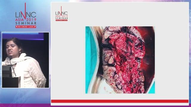
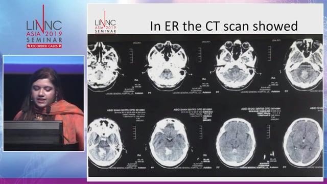
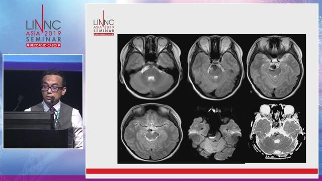
The first case was presented by Dr M. Noor Ul Huda (Pakistan). This case involved an infratentorial ruptured AVM with a prenidal aneurysm that was embolized with Onyx. This occluded both the nidus and the proximal aneurysm, without major deficit.
The debate that followed with the LINNC faculty members mainly concerned the origin of the hemorrhage itself, whether it was the nidus or the arterial aneurysm. In this case, which involved a right side hematoma and left paramedian nidus, the cause of the hemorrhage was seen to be the proximal aneurysm.
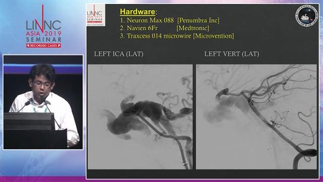
The second “attendee” case was presented by Dr Agarwal from India. The discussion that followed did not really focus on the modalities of treating this proximal A1 saccular aneurysm (coils only +/- remodeling balloon, which was seen as a good option and discussed by Profs. Spelle and In Sup Choi), but rather concerned the possibility of how to retrieve a stent that was not very well deployed.
Here, in this particular case, retrieval was successfully performed with a stentriever.
Recorded cases from Bicêtre
Case 1 from Bicêtre Hospital in France was presented by Prof. Jacques Moret. This case concerned a cavernous dural fistula with transmedian dural arterial supply and an ascending pharyngeal artery, without the possibility of transarterial access.
The technical choice was made to use a transvenous approach through the inferior petrosal sinus to obliterate the left cavernous sinus (using a jugular sheath, vertebral 5 Fr catheter, coils and Onyx).
Follow-up showed regression of symptoms and complete cure of the dural arteriovenous shunt on DSA. Audience discussion focused on the possibility of curing this dural shunt using only the coronal sinus without extending this to the entire left cavernous sinus.
Case 2 was presented by Prof. Spelle and involved multiple unruptured MCA aneurysms treated by WEB. This was a nice and detailed overview of why and how to choose the size of the WEB, and when to use an additional remodeling balloon. During the subsequent audience discussion, Prof. Spelle reminded us about the non-thrombogenicity of the tail of the WEB. He also spoke about how to react in case of perforation (don’t retrieve the WEB, prepare a balloon, consider deployment of the WEB and or additional coiling).
An industry sponsored symposium by Stryker followed in which Dr W. Lee from Singapore spoke about techniques for optimizing one pass TICI 3 in stroke.
Additional recorded cases from Bicêtre
The third recorded case of the day was presented by Prof. Laurent Spelle and involved a small, trilobulated ruptured Acom aneurysm. A transmedian remodeling balloon was used to help in the coiling of the aneurysm, with successive positioning of the microcatheter. A discussion followed on the necessity of preserving the communicating artery as well as the difficulties of accessing the microaneurysm related to the balloon catheter.
Industry sponsored symposium by Microvention provided a large overview by Prof. Jacques Moret about the WEB device. He explained how he categorizes the quality of WEB deployment, which depends on the complete or partial fitting of the WEB to the aneurysm. He also demonstrated the related long-term results related to this categorization. SLS seems to have better results on late follow-up. Other concepts were discussed, such as intentional protrusion of the WEB to get better exclusion of the aneurysm on follow-up. Concavity and long-term stability of the residual aneurysm after 18 months was well described. Using an additional stent in case of non-intentional and excessive protrusion of the WEB was described. We were warned of how we need to be careful of modifying the angulation of the parent artery after detachment, especially when using microcatheters larger than 17.
An Afternoon of cases and achievements
The afternoon began with the 4th recorded case from Bicêtre in which Prof. Moret showed us a frontal micro-AVM. This AVM was indicated by hemorrhage and had previously been treated by two successive embolisations with Glubran 2, leaving a small and not well defined residual nidus, with deep extension. Prof. Moret went on to describe all the technical issues involved in a combined transarterial and transvenous approach (a supra-selective arterial series to better define the arterial and venous angioarchitecture of the AVM, transarterial injection first, secondary transvenous injection, when to stop injecting in the venous side if you see a rupture in the subarachnoid non-hemorrhagic space). A Squid 12 proved to be especially good at filling the nidus.
Case 5 was of a 34-years-old patient with a left MCA stroke, admitted the day after onset to the hospital. Perfusion was very helpful in demonstrating and quantifying a mismatch (>1.8) between the MCA territory and the infarct, which was seen to be limited to the insular and front opercular area thus showing significant brain to save! Thrombectomy was successful (TICI 2b), performed using a solitaire stentriever and proximal balloon catheter. A carotid bulbar diaphragm was secondarily treated using a self-expanding stent (WEB). An iliac artery complication with perforation was treated by a covered stent (with the caveat of not using a covered stent more distally at the femoral artery level, at the place of flexion. If perforation had occurred during the first intervention at the acute phase of the stroke, it is advised to use glue in order to avoid double antiplatelet therapy with its risk of brain hemorrhage). The interactive discussion that followed revolved around the idea of the differential diagnosis between ruptured plaque and a carotid bulbar diaphragm.
An industry sponsored symposium by Medtronic entitled “How to maximize patient outcomes with your stroke portfolio offering” was presented by Adam S. Arthur from the USA. Concerned the fact that many patients arrive too late at the hospital, it is clear we need to improve the organization upstream of the time of the thrombectomy itself! We should be aware that for the family or patient, there is not the overriding fear of imminent death with a stroke, as we feel with a heart attack. And, except for the possible aphasia, it is important that we make the patient and family understand the true level of this emergency. This might account, in part, for the fact that 52% of thrombectomies are performed during the night or weekend.
An industry sponsored symposium by Siemens with Prof. René Chapot (Germany) looked at the new icono Biplane System. The quality of DSA is still technically moving forward!
The 6th recorded case from Bicêtre involved a 68-year-old woman presenting with right trigeminal pain which revealed an AVM of the pontocerebellum angle with a venous pouch of the draining vein compressing the fifth nerve, but without hemorrhage. Super selective catheterization of the AICA was first performed in order to better understand the supply of the AVM, but this is not the first choice for embolisation because of the labyrinthic artery at this level. Phil embolisation through the superior cerebellar artery was then performed. Prof. Moret demonstrated the use of a 3D roadmap used during the embolisation to control the progression of the embolic material through the entire nidus and the foot of the vein. A decrease of pain was observed. Audience discussion looked at regional arterial anatomies and the differential diagnosis as dural fistula.
Case 7 was of a wide neck anterior communicating aneurysm presented by Prof. Spelle. This was a very long and technically challenging intervention.
Using this recorded case, we were able to learn about the difficulty in trying to navigate a microcatheter VIA 33 to the anterior communicating region (compared to basilar tip or MCA bifurcation). The option of using a WEB device was abandoned. Failing deployment of a laser cut stent - along with the necessity of retrieving any other crossing microcatheter in order to retrieve the not completely deployed stent - offered further complications. Y-stenting was finally performed with two laser cut stents. VasoCT was used to clearly demonstrate the position of the two crossing stents, as well as the jailed microcatheter before coiling - this is the rule before the further steps which consist in the coiling of the aneurysm. The post-operative CT showed a subarachnoid hemorrhage, without the possibility of defining the exact point of bleeding. Repeated XperCT 30 mn and at 4 hours showed an increase of SAH. Antiplatelet therapy was stopped and nimodipine was administered to prevent vasospasm. A CT 24 hours later demonstrated a stabilized SAH and allowed for reintroduction of the antiplatelet treatment. In the end, the patient recovered well.
Best cases submitted by attendees
Four cases were awarded the coveted “best cases” submitted by attendees and, above all, underline the importance of interactivity between participants and faculty for our meeting.
The four chosen cases were:
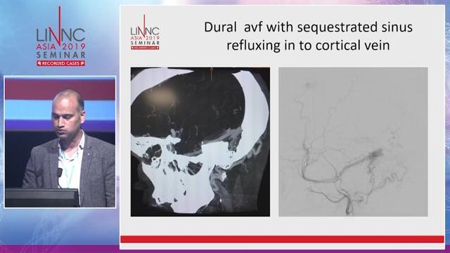
A case of a dural AVF with occluded lateral sigmoid sinus illustrating that recanalization of the sinus with transvenous approach was a good technical option.
A case of an intracranial multiple supra and infratentorial pial high flow fistulas, revealed by seizures in a 9-year-old boy.
The technical options for treatment were a transarterial approach using coils and glue. No HHT history, and typical phenotypic aspects.
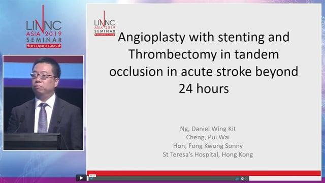
A case of multiple intracranial atherosclerotic stenoses (right ICA, left A1), presenting with a right hemispheric haemodynamic stroke. After failure of optimized medical therapy and recurrence of the stoke, a left A1 angioplasty was performed, followed by stenting of the left A1 using a laser cut stent.
This case inspired an interesting discussion on why not to use a coronary stent, due to unfavorable anatomy (the curve of the A1 not being compatible with the rigidity of coronary stent).
We also discussed an MCA bypass and the morbidity linked to surgery.
A case involving a dAVF around the anterior condylar confluent which offered a nice anatomical review of the condylar veins and an original therapeutic approach through the paraspinal venous plexus.
Stay tuned for Day 2!
And don’t forget to follow our live coverage of #LINNCASIA on
Facebook
,
Twitter
and
Linkedin
!
Reported by :
|
|
|
| Augustin Ozanne and Sophie Gallas
|
Read the report from Day 2