CASE PRESENTATION
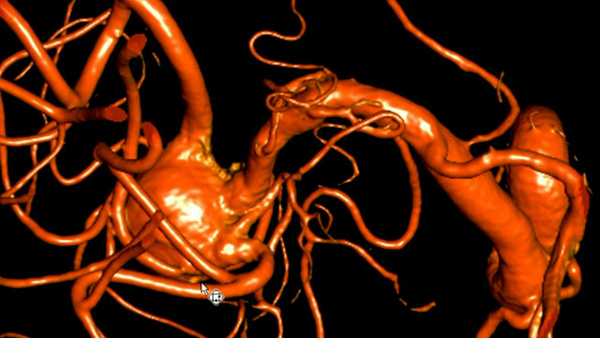
Early 50s female patient with Hunt & Hess Grade 5 subarachnoid hemorrhage was found to have a ruptured right middle cerebral artery (MCA) bifurcation aneurysm which encased the superior division at its base.
The aneurysm dome showed two blebs on it. The aneurysm measures 8.3mm transverse, 5.4mm craniocaudal, and 7.2mm anterior-posterior dimensions with the neck size of 3.2mm.
Fig. 1. Right MCA aneurysm with acute M1 spasm
Fig. 2. Right MCA aneurysm with M1 spasm magnified view
TREATMENT APPROACH
Due to the grade and acute spasms, endovascular intervention was chosen over open surgery.
Due to the irregular shape and branch encasement, double microcatheter technique was utilized.
SET UP
A 6 French guide sheath was placed in the upper cervical internal carotid artery.
A Hyperglide 4x15mm balloon was prepped in case of rupture or a need to convert to balloon assisted embolization, but not advanced into the vasculature.
An Echelon 10 micro-catheter with 45 pre-shaped tip was advanced into the MCA over Xpedion 10 guide-wires.
3mg of intra-arterial Papaverine was slowly infused into the MCA M1 segment via the micro-catheter prior to advancing it into the aneurysm.
Once the micro-catheter was advanced into the aneurysm, a Target 360 ultra 3x8 was deployed as a framer, which was intentional undersized to avoid herniation of the coil to the parent artery or the encased superior division.
Next, another Echelon 10 micro-catheter with straight tip was advanced into the aneurysm and additional Target 360 ultra 3x6 was slowly inserted to the aneurysm with the initial coil undetected.
The intention for using two different tip shape was to deliver the coils to the different directions in the aneurysm.
Once the two coils were tangled together conforming to the irregular shape of the aneurysm dome, the frame became much more stable.
The initial coil was detached and additional coils were deployed into the aneurysm via the two micro-catheters.
A total of 6 coils (23cm) were deployed into the aneurysm.
There was a residual aneurysm neck, but no aneurysm dome was opacified.
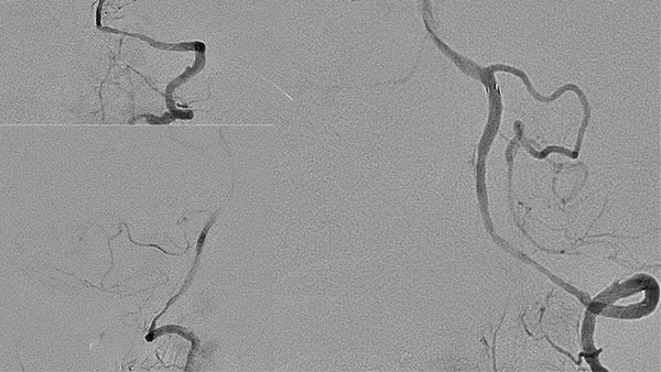
Fig. 3. The 1st micro-catheter placed in the aneurysm dome following intra-arterial 3mg Papaverine injection
into the spasmed M1.
Fig. 4. Initial framing coil in the aneurysm dome.
Fig. 5. 2nd framing coil was deployed. With two coils,
the frame structure became very stable in the aneurysm.
Fig. 6. Additional coils deployed into the frame via the micro-catheters
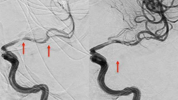
Fig. 7. Final angiogram shows no residual dome filling
with preservation of the encased superior division of MCA.
POST PROCEDURE
The patient condition did not changed after the intervention.