- 69-year-old female patient
- Arterial hypertension (AHT)
- No other diseases
- NIHSS: 18
- Weakness of the right limbs and expressive aphasia
- Onset time: 11.00 am
- Arrival at the hospital: 12.24 pm
Neuroimaging - CT: aspect 7
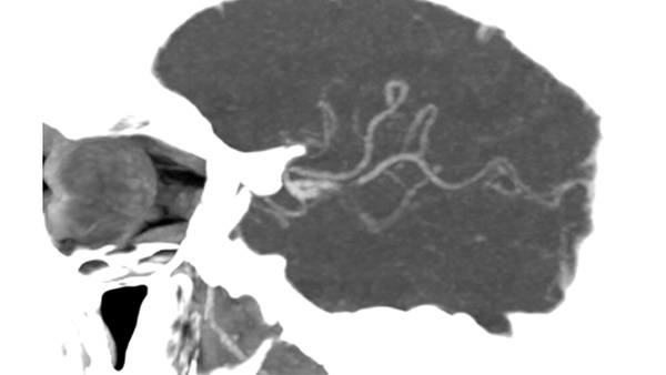
AngioCT: M2 left occlusion
AngioCT: M2 left occlusion
Perfusion: Even though the perfusion parameters are not favorable, we ignored this information due to the time of evolution since the beginning of the clinic.
Thrombectomy
- Arrival into angiosuite: 13.15
- Time of puncture: 13.33
- First imaging: 13.52
- Recanalization: 14.21
Treatment with alteplase (bolus + start of perfusion) is indicated and MT is performed, which must be performed under sedation due to intense agitation (13 mg of Midazolam was required).
Devices Used: Double Solitaire
- Parietal Branch: Phenom 21, Traxcess, Solitaire 6x40
- Silvian Branch: SL10, Traxcess, Solitaire 3x20
- Flow gate 2
- Final result TICI 2C after 1 pass
In our experience, the use of first-intention stentrievers in acute ischemic stroke is safe and effective. In patients with two or more affected branches, it is possible to act on both occlusions in the same pass.
Imaging – Pre MT
You do not have permission to view this object.
You do not have permission to view this object.
Imaging – Pre MT
You do not have permission to view this object.
Double stenting technique where both stents are removed at the same time
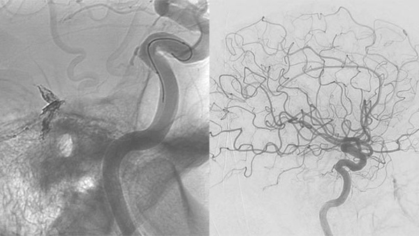
Imaging – post MT
You do not have permission to view this object.
You do not have permission to view this object.
Comparison of pre and post MT imaging
Post MT
- The patient was admitted to ICU for clinical surveillance and early extubation.
- Very significant clinical improvement to NIHSS = 0 after 24 hours were observed and patient started rehabilitation.
- 24-hour control cranial CT showed a slight sylvian petechial bleed which wasn’t symptomatic.
- Simple antiplatelet therapy was started with clopidogrel.
- Patient was discharged from hospital 9 days post-MT and was neurologically asymptomatic.
CT 24-hour post-MT