A 44-year-old man was referred to the neurointerventionalist six hours after sustaining a shotgun wound to the left chest, shoulder, and neck from four feet.
Physical examination of the chest showed a 5 (cm) x 5 (cm) gunshot entry wound on the anterior-superior aspect of the chest involving the supra and infra-clavicular region, with multiple gunshot pellet entry sites riddled in the surrounding vicinity.
The patient was taken for a computed tomography (CT) scan of the brain without contrast and a computed tomography angiography (CTA), that showed no sign of stroke or intracranial hemorrhage, but revealed a single “buckshot” pellet embolizing the basilar artery tip partially occluding the origin of the left posterior cerebral artery (PCA).
A Direct Aspiration First Pass Technique (ADAPT) was used to pull back the shotgun pellet to the V1 segment of the right vertebral artery (RVA) where it got stuck at the tip of the Neuron MAX guide catheter, and through continuous aspiration, the Neuron MAX guide catheter was pulled along with pellet.
Through this technique, the neurointerventinalist was successfully able to endovascularly remove the embolized pellet, and the patient was discharged 8 days later with no focal neurological deficit.
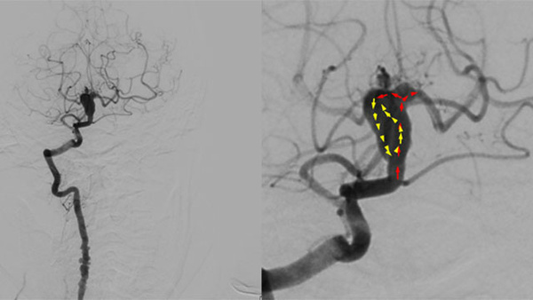
Figure 1:
- Figure 1.A: Digital Subtraction Angiography frontal view showing shotgun pellet at the top of the basilar artery partially occluding the origin of the left posterior cerebral artery (PCA).
- Figure 1.B: Digital Subtraction Angiography lateral view showing shotgun pellet at the top of the basilar artery partially occluding the origin of the left posterior cerebral artery (PCA).
- Figure 1.C: Digital Subtraction Angiography frontal view showing shotgun pellet at the top of the basilar artery partially occluding the origin of the left posterior cerebral artery (PCA) with measurements of the shotgun pellet diameter (2.5 ± 0.33 (mm)), right PCA diameter (2.1 ± 0.33 (mm)), left PCA diameter (1.8± 0.33 (mm)), and the vertebrobasilar artery diameter (2.8 ± 0.33 (mm)).
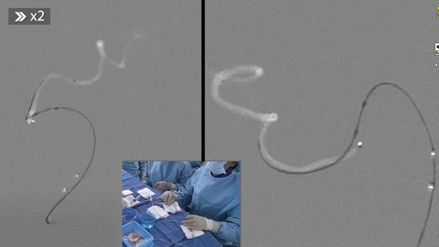
Figure 2:
- Figure 2.A, 2.B: Shotgun pellet stuck on the tip of the guide catheter.
- Figure 2.C: Shotgun pellet once removed from the tip of the guide catheter exhibiting no physical evidence of thrombus.
Watch the video narrated by Ameer E. Hassan :
- Direct Aspiration First Pass Technique (ADAPT), used to pull back shotgun pellet to V1 segment of the right vertebral artery (RVA) where it got stuck at the tip of the Neuron MAX guide catheter, and through continuous aspiration Neuron MAX guide catheter was pulled along with pellet.
You do not have permission to view this object.
Reference :
https://jnis.bmj.com/content/12/2/e2