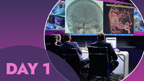
- Day 1 at LINNC Seminar 2018, Asia Edition -
Good morning and welcome to the LINNC Seminar 2018, Asia edition in Singapore.
We’ve traveled across the world to share our experience and help define INR techniques and strategies in the setting of neurovascular pathologies. Together, we can improve what we do each day by exchanging knowledge, allowing our colleagues – and ourselves – to work more efficiently, effectively and safely. The recorded cases that we saw today (and will see tomorrow) further reinforce the message that our results and our experience – as well as our mistakes – when they are shared freely and equally in the open and trusting environment of a LINNC seminar, can create a greater cohesion between all INR specialists.
The cases begin
The first case was an extradural arteriovenous fistula presented by Junjie WANG. This was very interesting as a combined approach was used in the setting of a difficult and recurrent epidural fistula. The key message here was that we need to understand the pathology.
Then Alvin Yichou Wang presented a recorded angioplasty case in which two wires were used, each in both branches of a bifurcation. At the present time, we need more studies concerning this approach and are not sure if it is better than angioplasty with stents. The patient follow-up is now at 3 years, but a certain rate of restenosis and complications can occur. This led to discussion concerning the setting of intracranial stenosis.

After this, Prof. Jacques Moret from Bicêtre Hospital, showed us a recorded case of a vein of galen AVM treated in a 4-year-old girl. A truly wonderful technical presentation with one clear message: "now, using a detachable catheter – even with glue – you have time!". Eight minutes for injecting the glue, with continued progression. Many techniques could be employed here, such as using a balloon or coiling first, but slow injection is a simple way of achieving your goal. These are just a few of the ideas that Prof. Moret challenged us with in his opening case today, spicing up the discussion and putting a bit of fire in the room. Many physicians argued their own points-of-view on this subject, as well as answering a variety of questions from the audience… all of us present following the debate with great interest and attention.
We then turned to Prof. Laurent Spelle who presented a case of a thrombosed intracavernous aneurysm where the main technical challenge was to deliver not one but two flow diverters through a Copernic balloon with a double lumen.
Industry partners
After a coffee break, the Stryker symposium was presented by M.J. Gounis in which we thoroughly explored the different techniques that can be used to maximize stroke treatment. This was a great presentation, that offered results from in vitro research recalling the clinical benefits of maximizing thrombectomy techniques.
Return to cases
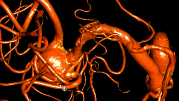
Prof. Moret presented the next recorded case, looking at a small basilar tip aneurysm with a very irregular shape, a clinical situation that is very challenging to treat. Hopefully, today we have arrived at that optimal point where we can properly analyze the anatomy involved in these situations and choose the best adapted devices.
Prof. Spelle then showed us a case concerning Y configuration stent-assisted coil embolizations using laser-cut open-cell stents. While this is technically feasible, the faculty discussed different protocols for treating this disease, from surgical to interventional treatments.
Back to industry
The first session ended with a Medtronic symposium looking at the endovascular treatment for acute stroke associated with intracranial atherosclerotic stenosis. A pathology with no clearly defined guidelines, the challenge was to generate faculty interest with the door open to further research.
Lunch break was very welcome after these intense discussions and such a full, first morning.
The afternoon begins
We started with a recorded case from Bicêtre Hospital of the endovascular treatment of a parietal AVM. The classic endovascular approach is to use a transarterial access with the selective catheterization of an arterial feeder as close as possible to the nidus. Nevertheless, when arterial access is not possible and other treatment methods are not applicable or carry too high a risk for the patient, the transvenous approach is a very good option. The technique for venous catheterization is different however, and we might face difficulties in navigating retrogradely into the target vein. Our experience, especially our failures in this area are further amplified, and become more meaningful and understood if we can share them.
We then saw cases of stroke treatment using an EmboTrap, where TICI 3 was achieved after one pass.
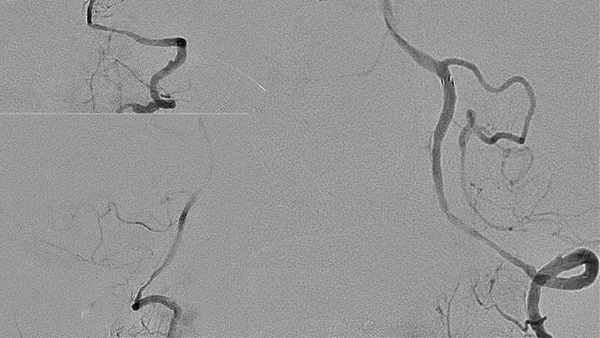
A recorded case with a Pipeline stent placed in front of a large PComA aneurysm offered a very interesting illustration of how flow velocities change after stent deployment using mean aneurysm flow amplitude (MAFA). Occlusion of the aneurysm after FD stenting is determined by multiple factors including aneurysm size, side branches and the hemodynamic environment. The MAFA-R is an available tool to predict aneurysm occlusion.
The Microvention symposium presented by A. Boulos concerned hydrocoils.
A short break, and then…
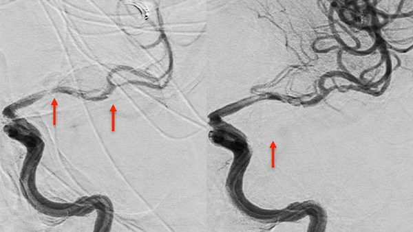
After the coffee break, two recorded cases were presented. The second was a case of a giant dissecting M1 aneurysm. Despite remarkable recent advances in the technical and procedural success of endovascular therapy, parent vessel occlusion is still an option in certain cases. This case highlighted the importance of collateral vessel status.
Finally, presentations of select cases submitted by attendees were shown. Dr. Lin from Taiwan introduced transvenous embolization of dural CCF via pterygoid plexus; Dr. Ren from Beijing, China, showed us a patch embolization for a blister-like aneurysm, and from India, Dr. Das showed us a complex fusisaccular MCA aneurysm treatment with FD. All interesting, understandable and highly teachable work.
Until tomorrow
A long day, with lots of information and discussion, but it's not over yet. We will see you tomorrow for the last day of LINNC Seminar 2018, Asia edition in Singapore…
Reported by
Vanessa Chalumeau
Read the day 2 report