- A 54-year-old patient arrives at the emergency room due to a sudden-onset aphasia and right-sided hemiparesis
Brain MRI, performed under the strong suspicion of a stroke, showed:
- Acute ischemic lesion in the left fronto-parieto-temporal region
- Subarachnoid hemorrhage predominantly located in the left Sylvian fissure
- Two cerebral aneurysms at the bifurcation of the M1 segment and at the bifurcation of the inferior M2 segment of the left MCA
- Severe vasospasm of left MCA
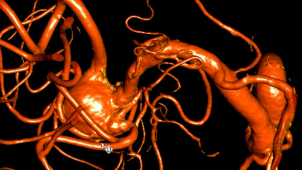
DSA
Two wide-neck aneurysms with a very irregular shape, moderate vasospasm of the M1 segment and severe vasospasm of the M2 branches of the left MCA.
Main concerns
- Management of two aneurysms during the same session
- Wide neck
- Very irregular shape
- Highly unfavorable attack angle for accessing the more proximal aneurysm
- Severe vasospasm
The multidisciplinary discussion highlighted the need to rapidly secure the aneurysms and the impossibility of performing neurosurgery due to severe vasospasm.
Therapeutic strategy
- Management of vasospasm followed by endovascular treatment of both MCA aneurysms.
Endovascular procedure
- Right femoral arterial access was obtained using an 8F short introducer.
- A Neuron Max guiding catheter was positioned at the level of the left carotid bulb.
- A Sofia 6 intermediate catheter was positioned in the left internal carotid artery.
First step: vasospasm management
Administration of vasodilators: 3 mg of Nimodipine followed by 2 injections of 5 mg of Milrinone) until the arterial caliber was sufficient to allow the passage of the microcatheters, allowing endovascular procedure.
3D acquisition post-vasospasm treatment allows a more detailed analysis of the aneurysms in order to plan the intervention.
Both lesions present an irregular-shaped sac and a wide neck.
Therapeutic strategy: treatment of both aneurysms using WEB™ devices
Endovascular treatment of the 1st distal aneurysm
- Selective microcatheterization of the inferior M2 segment bifurcation aneurysm was performed using a VIA 17 microcatheter over a Terumo 0.012" microguidewire with a 90° tip.
- A WEB™ SL 4 mm x 3 mm device was deployed in the aneurysmal sac and subsequently detached.
Endovascular treatment of the 2nd proximal aneurysm (same session)
- Selective microcatheterization of the M1 segment bifurcation aneurysm was performed using the same VIA 17 microcatheter over a Terumo 0.012" microguidewire.
- A WEB™ SL 3.5 mm x 2 mm device was deployed within the aneurysmal sac and subsequently detached.
DSA after endovascular treatment
Correct deployment of both WEB. Contrast medium stagnation within both aneurysms.
24-hour DSA
The 24-hour angiography showed an early adequate occlusion with the presence of neck remnant for both aneurysms (yellow arrows for the proximal, blue arrows for the distal).
In the following days, the patient underwent multiple endovascular treatments for cerebral vasospasm.
The patient was discharged from the hospital approximately three weeks after the acute event and was referred to a rehabilitation program due to speech impairment and right-sided motor weakness resulting from the ischemic lesion caused by vasospasm at the moment of the diagnosis.
6-month follow-up
The 6-month DSA showed the complete occlusion of the distal aneurysm, and the persistence of a small neck remnant of the proximal aneurysm (arrows).
18-month follow-up
Conclusion
- Cerebral vasospasm after SAH is a common complication that occurs in up to 70% of patients. While the most common time for vasospasm to develop is 3-7 days post-SAH, some patients experience it within the first 24 hours.
- Early and severe vasospasm can result in new focal neurological deficits, decreased level of consciousness, or other symptoms like weakness, sensory changes, and headache.
- Severe cerebral vasospasm after subarachnoid hemorrhage (SAH) is not an absolute contraindication for surgical clipping, but some studies suggest it might be associated with less vasospasm compared to surgical clipping.
- The WEB device is safe and effective in treatment of ruptured bifurcation aneurysms.
- In this challenging case of SAH, with an atypical presentation, double aneurysms and severe early vasospasme, the WEB™ Embolization System allowed the prompt treatment of two cerebral aneurysms with unfavorable anatomy, ensuring rapid securing in the acute phase and an excellent long-term outcome.
References
- Jabbarli R, Reinhard M, Shah M, Roelz R, et al. Early Vasospasm after Aneurysmal Subarachnoid Hemorrhage Predicts the Occurrence and Severity of Symptomatic Vasospasm and Delayed Cerebral Ischemia. Cerebrovasc Dis. 2016;41(5-6):265-72. doi: 10.1159/000443744.
- Imamura H, Tani S, Adachi H, Fukumitsu R, Sunohara T, Fukui N, Omura Y, Sasaki N, Akiyama T, Fukuda T, Kajiura S, Shigeyasu M, Asakura K, Horii R, Sakai N. Comparison of Symptomatic Vasospasm after Surgical Clipping and Endovascular Coiling. Neurol Med Chir (Tokyo). 2022 May 15;62(5):223-230. doi: 10.2176/jns-nmc.2021-0126. Epub 2022 Apr 12. PMID: 35418528; PMCID: PMC9178112.
- Spelle L, Herbreteau D, Caroff J, et al. Clinical Assessment of WEB device in Ruptured aneurYSms (CLARYS): results of 1-month and 1-year assessment of rebleeding protection and clinical safety in a multicenter study. J Neurointerv Surg. 2022 Aug;14(8):807-814. doi: 10.1136/neurintsurg-2021-017416.
Information and/or case images provided may not represent the approved indication for use for each country/market. Please refer to the Instruction for Use (IFU) in the specific market/country that you are looking into. Intended for Healthcare Professionals in EMEA Only. Legal Manufacturer: MicroVention, Inc. / EU Authorized Representative: MicroVention Europe S.A.R.L. WW
Back to 2025 Clinical Case Contest